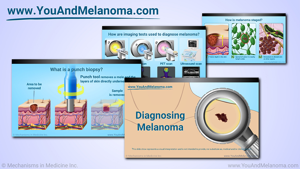
Diagnosing Melanoma
*Please note: This slide show represents a visual interpretation and is not intended to provide, nor substitute as, medical and/or clinical advice.
How is melanoma diagnosed?
Melanoma is usually diagnosed by a dermatologist, a doctor who specializes in skin diseases.
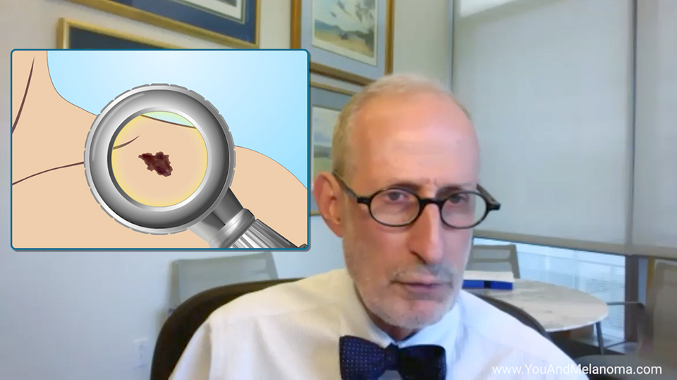
Your doctor should carefully examine your skin - including the more private areas - to look for unusual changes.
Your doctor will assess the size, shape, color, and texture of all spots on your skin, especially any “ugly ducklings” that look different from their neighbors.
What is a biopsy?
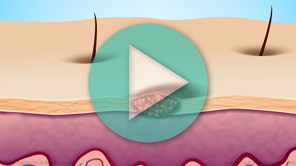
Your doctor may do a biopsy of any suspicious-looking mole. They will remove all or part of the mole and send it to a lab, where it will be looked at under a microscope.
What is a punch biopsy?
In a punch biopsy, the doctor uses a pencil-like tool to remove a mole and the layers of skin directly underneath it.
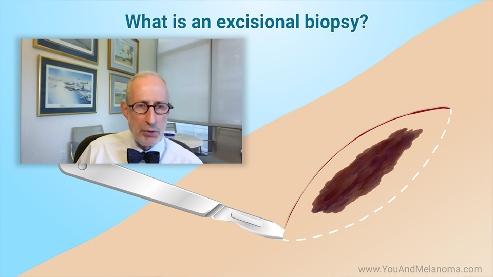
What is an excisional biopsy?
In an excisional biopsy, the doctor uses a small knife called a scalpel to remove the mole and some of the normal skin around and below it.
What will the pathology report tell you?
After the biopsy, you will get a detailed report of its results. This is called a pathology report. Some of the things it will tell you are:
-
Were cancer cells found? If so, what kind? And how far have they spread?
What will the pathology report tell you?
-
How deep is the tumor at its thickest point? This is called the Breslow depth.
- How deeply has the tumor penetrated the skin? This is called the Clark level. This is not the tumor stage - we'll get to that in a minute. Doctors no longer use the Clark level to predict a patient's outlook, but it is still usually included in the pathology report.
What will the pathology report tell you?
-
How close does the tumor come to the edge of the skin sample? This is called the margin.
-
How many cells in the skin sample are actively dividing? This is called the mitotic count or mitotic rate. A higher number means that more cells are dividing.
-
Is there ulceration? This is when there is a separation of the outer and inner layers of skin and may be associated with bleeding.
What are lymph nodes?
Lymph nodes are part of your immune system. Hundreds of them are located throughout your body, with many found in the neck, armpits, elbows, and groin.
Lymph nodes are small organs that contain and store large numbers of lymphocytes, white blood cells that help the body fight infection and diseases like cancer.
What is a lymph node biopsy?
A
lymph node biopsy
is done to find out if melanoma has spread from the skin to nearby lymph nodes. All or part of a lymph node is removed and sent to a lab to be looked at under a microscope.
What are the types of lymph node biopsy?
In a fine needle aspiration biopsy, the doctor uses a syringe with a thin, hollow needle to remove tiny pieces of a lymph node.
What are the types of lymph node biopsy?
In a sentinel lymph node biopsy, the doctor injects blue dye into the area around the tumor. The lymph nodes that get stained with dye are the ones that melanoma is likely to spread to first. The dye-stained lymph nodes are removed and tested for cancer.
What are the types of lymph node biopsy?
In an excisional lymph node biopsy, the doctor makes a small cut in the skin to remove an enlarged lymph node. The node is then tested for cancer.
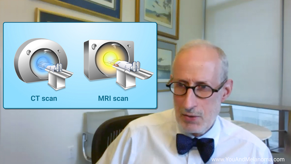
How are imaging tests used to diagnose melanoma?
Imaging tests may be done to look for cancer cells throughout the body.
-
An MRI scan uses radio waves and magnets to create detailed pictures of areas inside the body.
-
A CT scan uses a computer linked to an x-ray machine to make 3D pictures of areas inside the body.
-
In a PET scan, you will be given an injection of a small amount of radioactive sugar. A special camera makes detailed pictures of the areas inside the body where the sugar collects. Cancer cells absorb much more of the sugar than normal cells do.
- An ultrasound scan uses sound waves to form pictures of tissues and organs on a computer screen.
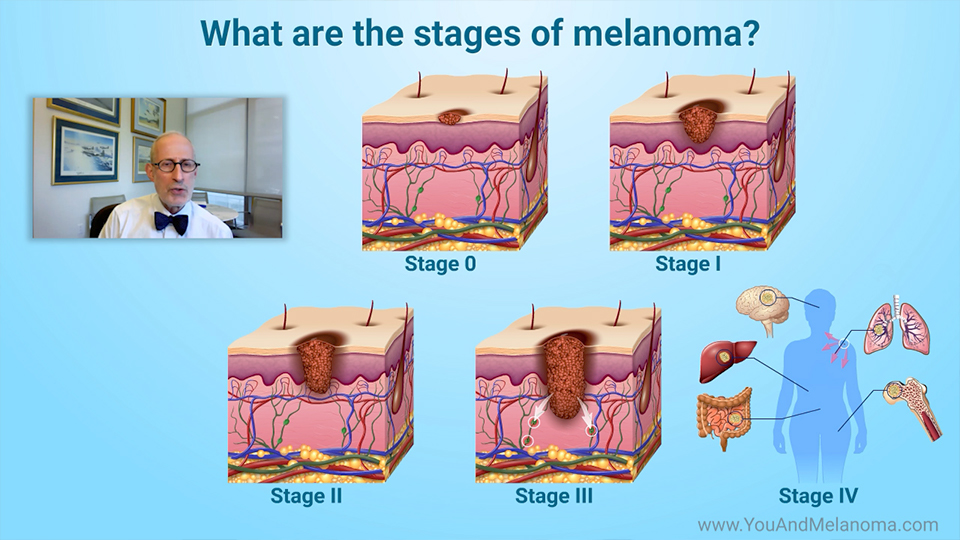
How is melanoma staged? - TNM
Staging is a way of describing how much cancer is in the body and how far it has spread. Doctors do this using a system known as TNM, which can predict a patient's outlook.
-
T is for tumor depth in the skin.
-
N is for spread of the cancer to nearby lymph nodes.
-
M is for metastasis, or spread of the cancer to distant skin or other organs.
How is melanoma staged? – Stage 0
Doctors combine the T, N, and M to get an overall cancer stage.
-
Stage 0 melanoma is limited to the top layer of the skin. This is also known as melanoma in situ. The chance that it will spread is extremely low.
How is melanoma staged? – Stage I and II
-
Stage I melanoma is limited to the skin and is at most 2 mm deep.
- Stage II melanoma is limited to the skin and is more than 2 mm deep.
How is melanoma staged? – Stage III
- Stage III melanoma may be any depth, and the cancer cells have spread to nearby skin or lymph nodes.
How is melanoma staged? – Stage IV
-
Stage IV melanoma is metastatic. The tumor may be any depth, and the cancer cells have spread to distant lymph nodes or organs, such as the lungs, liver, brain, bones, and intestines.
References
- Melanoma Skin Cancer Stages. American Cancer Society. https://www.cancer.org/cancer/melanoma-skin-cancer/detection-diagnosis-staging/melanoma-skin-cancer-stages.html
- Melanoma: Stages. American Society of Clinical Oncology. https://www.cancer.net/cancer-types/melanoma/stages
- NCCN Guidelines for Patients. National Comprehensive Cancer Network. https://www.nccn.org/patients/guidelines/cancers.aspx
- NCI Dictionary of Cancer Terms. https://www.cancer.gov/publications/dictionaries/cancer-terms
- Tests for Melanoma Skin Cancer. American Cancer Society. https://www.cancer.org/cancer/melanoma-skin-cancer/detection-diagnosis-staging/how-diagnosed.html
- Urso C. Are growth phases exclusive to cutaneous melanoma? J Clin Pathol 2004 May;57(5):560.
This slide show describes the tests and procedures doctors use to
diagnose melanoma. It explains skin examination, skin
biopsy methods (punch biopsy, excisional biopsy)
lymph node biopsy methods (fine needle aspiration biopsy, sentinel lymph node biopsy, excisional lymph node biopsy), and
imaging tests (MRI, CT scan, PET scan, ultrasound). This slide show also provides an overview of how melanoma is
staged (how far the cancer has spread). Knowing the stage helps doctors decide how to best treat your melanoma and predict your
prognosis, or outlook.
-
Share with family and friends:
Click here to take our SURVEY
Your feedback is important to us! We will use your feedback to develop future areas of content about melanoma which will help other patients, caregivers, and families.